CAT Scanning
Saint Paul Goes to the Hospital
The Original Question
NG3899 (figure 1) is a small painting depicting Saint Paul by an artist active in Milan in the 1490s and early 1500s known as the Master of the Pala Sforzesca. It was once part of a larger structure, probably an altarpiece. During recent technical examinations the current painting support was seen to be composed of two separate wooden panels. The original panel (type of wood not identified) was used with grain running horizontally. Stuck to the back of the original wood is a second panel (figure 2) the wood of which has been identified as walnut and which has grain running vertically.
A conventional x-ray image was made which, in addition to information about the paint layers, clearly shows three nails embedded somewhere in the panel structure, towards the bottom of the picture (figure 3). As the nails are not visible from the front or back of the panel the question was raised, is there a type of imaging that would show where in the structure the nails are positioned?
It was proposed that a CAT scan, which gives x-ray images in three dimensions, might provide the information desired.
| {{#iipimage:
server=http://research.ng-london.org.uk/fcgi-bin/iipsrvN.fcgi%7C image=/pics/pyramids/rwCode/ngl/ng3899/N-3899-00-000012-PYR.tif| wid=200px|hei=200px|format=left| caption= Figure 1: NG3899 Master of the Pala Sfozesca, Saint Paul. }} |
{{#iipimage:
server=http://research.ng-london.org.uk/fcgi-bin/iipsrvN.fcgi%7C image=/pics/pyramids/rwCode/ngl/ng3899/N-3899-00-000017-PYR.tif| wid=200px|hei=200px|format=left| caption=Figure 2: NG3899 Image of the back of the panel. }} |
{{#iipimage:
server=http://research.ng-london.org.uk/fcgi-bin/iipsrvN.fcgi%7C image=/pics/pyramids/rwCode/ngl/ng3899/N-3899-00-000014-PYR.tif| wid=200px|hei=200px|format=left| caption=Figure 3: NG3899 Original X-ray Image. }} |
Results
General Electric (GE) make the CAT scanners used in many hospitals and were only too pleased to provide their technology to help the National Gallery in this intriguing investigation.
The painting was taken to The Princess Grace Hospital where it was scanned using their High Resolution GE Discovery HD 750 (figures w,x,y,z) The results of the CAT scan consist of a series of x-ray images showing "slices" through the painting in each of three directions: coronal, sagittal and axial.
- Coronal images (figs 4 - 9) show slices through the painting parallel to the paint surface at different depths.
- Sagittal images (figure 10) show slices through from front to back of the panel parallel to the side edges.
- Axial images (figure 11) show slices through from front to back of the panel parallel to the bottom edge.
The painting was resting at a slight angle in the scanning bed, the feet of the saint slightly higher than the head, with the result that some of the coronal images show a combination of information from adjacent layers (see figs 4,5,& 7).
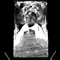
Figure 4: Coronal image 1, top slice, paint of top half of figure, with original wood (horizontal grain) in bottom half of image.
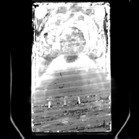
Figure 5: Coronal image 2, lower paint and original wood (horizontal grain), nails begin to show
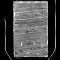
Figure 6: Coronal image 3, original wood with 3 nails
Figure 7: Coronal image 4, original wood and second panel (vertical grain)
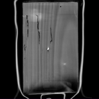
Figure 8: Coronal image 5, second panel, vertical wood grain
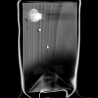
Figure 9: Coronal image 6, bottom slice, second panel with seal


Figure 6 clearly shows the three nails with the horizontal grain of the original wood and none of the second panel. Their position is further clarified by looking at axial scans (figure 11). The nails (the three white dots) are clearly within the upper (original) wood. They are not original but added as part of a repair before the second panel was attached.
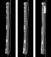
Figure 10: 3 Sagittal images each showing one nail, in the original wood.
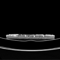
Figure 11: Axial image showing location of the three nails within the original wood.
Figure 12: Coronal and axial images combined.


Related Links
- National Gallery press release.
- http://www.nationalgallery.org.uk/paintings/master-of-the-pala-sforzesca-saint-paul
- High Resolution Images
- Further technical information about the painting has been published as part of the article: Spring, M., Mazzotta, A., Roy, A., Billinge, R., Peggie, D. 'Painting Practice in Milan in the 1490s: The Influence of Leonardo'. National Gallery Technical Bulletin Vol 32, pp 78–112.